
Learning Experience & E-Module Design
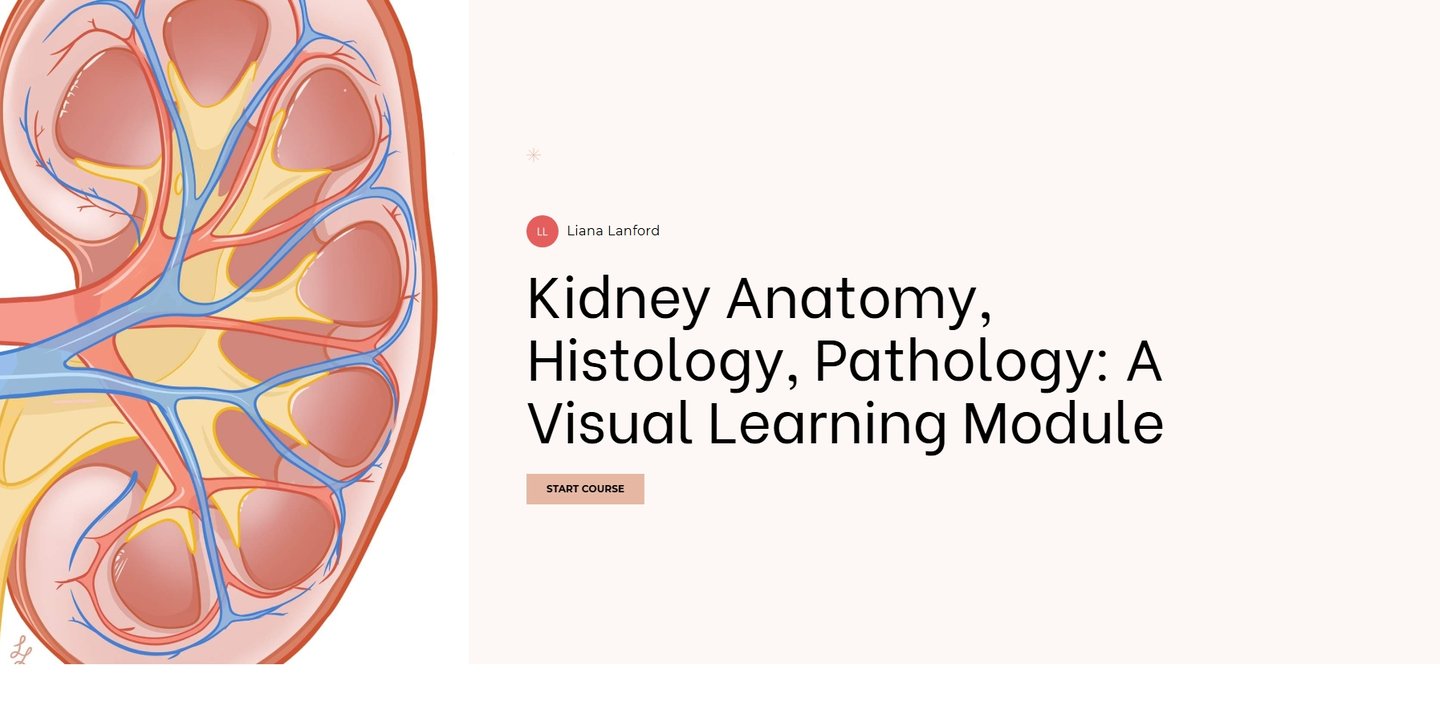
Kidney Anatomy, Histology, Pathology: A Visual E-learning Module Prototype
This master's research project focused on the design and development of an interactive e-learning module for teaching kidney anatomy, histology, and pathology to graduate and medical students. The prototype integrates 3D models, medical illustrations, histological imagery, animations, and interactive assessments into a unified educational experience. Guided by principles of instructional design and multimedia learning, the project aimed to improve learner engagement, support knowledge retention, and enhance understanding of complex biomedical concepts. By combining biomedical visualization, scientific communication, and educational technology, the module demonstrates how interactive learning experiences can improve the accessibility and effectiveness of healthcare education.
Created using Procreate, Adobe Illustrator, Cinema4D, Adobe Substance Painter, Articulate Rise 360.
Illustrations
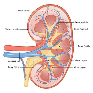
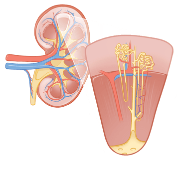

Inside cross-section of kidney
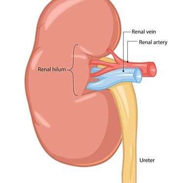
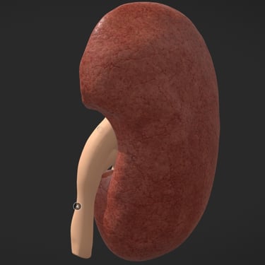
External kidney
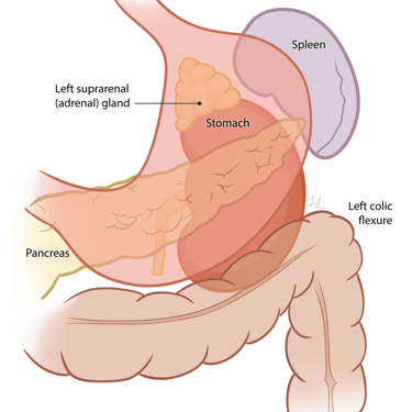
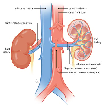
Relationship to surrounding structures (Left kidney)
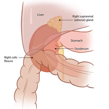
Relationship to surrounding structures (Right kidney)
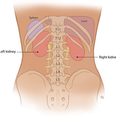
Position of the kidneys retroperitoneally
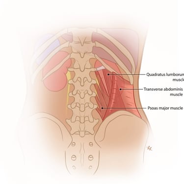
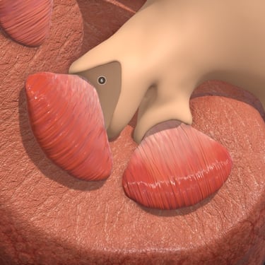
Muscles directly overlaying the kidney
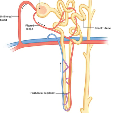
Cross-section & callout of nephron
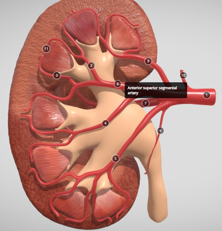
Blood supply of the kidneys
Labelled nephron of the kidney
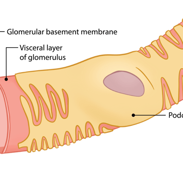
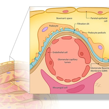
Inside of the Renal Corpuscle
Glomerular filtration barrier
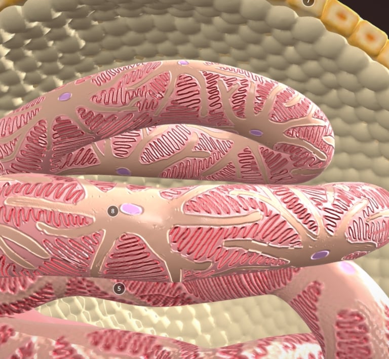
Cross-section of Renal Corpuscle cells
Illustrations created using Illustrator, labels created with Adobe Illustrator
3D Models
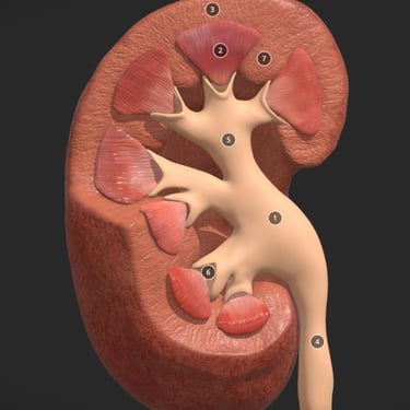
Kidney cross-section
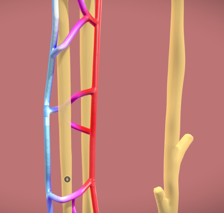
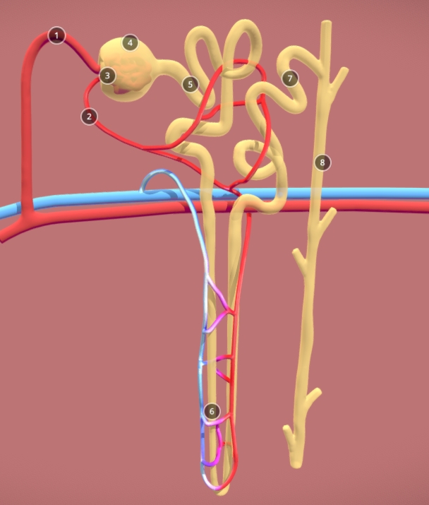
Functional unit of the kidney: the nephron
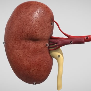
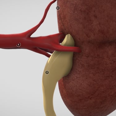
Simple external kidney with ureter, renal vein, and artery
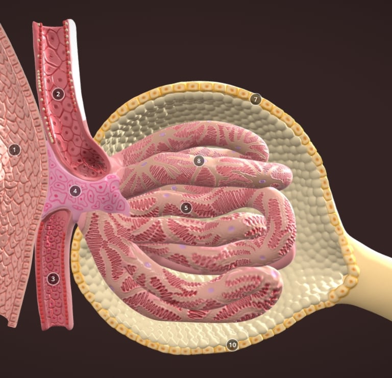
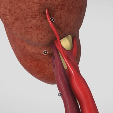
Renal corpuscle of the nephron cross-section
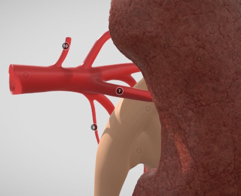
Renal artery branches of the kindey
Created using Cinema4D, Adobe Substance Painter
Contact
Reach out for commissions or collaborations
contact
Copyright Lanford Biomedical © 2026. All rights reserved.