
Medical Illustration
Aqueous Humor Dynamics and Glaucoma Anatomy
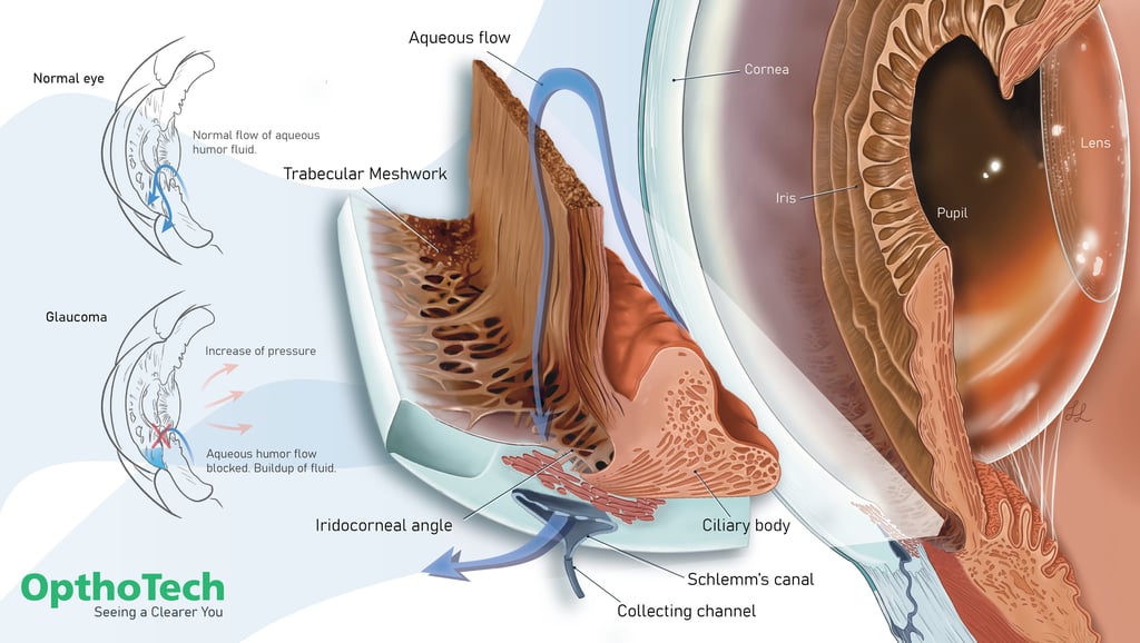
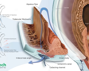
Detailed medical illustration of the anterior chamber of the eye, emphasizing the iridocorneal angle, trabecular meshwork, Schlemm’s canal, and the normal pathway of aqueous humor drainage. The composition also compares normal aqueous outflow with glaucomatous obstruction, illustrating how impaired drainage can lead to increased intraocular pressure. Created in 2026 using Procreate and Adobe Illustrator.
Aqueous Humor Dynamics and Glaucoma Anatomy

Detailed medical illustration of the anterior chamber of the eye, emphasizing the iridocorneal angle, trabecular meshwork, Schlemm’s canal, and the normal pathway of aqueous humor drainage. The composition also compares normal aqueous outflow with glaucomatous obstruction, illustrating how impaired drainage can lead to increased intraocular pressure.
Created in 2026 using Procreate and Adobe Illustrator.
Acute Compartment Syndrome: Anatomy and Pathophysiology
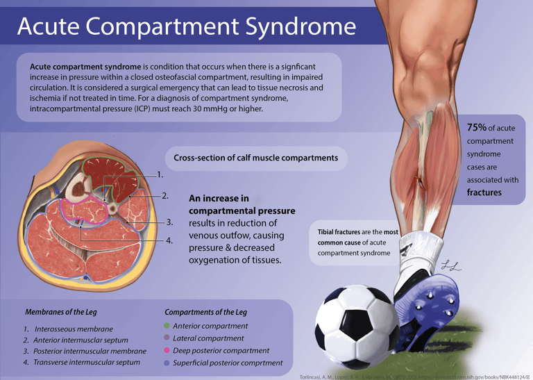
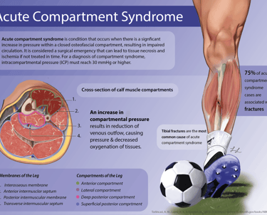
This educational medical illustration examines the anatomical and physiological basis of acute compartment syndrome within the lower leg. Through a detailed cross-sectional visualization of the calf compartments and supporting explanatory graphics, the poster demonstrates how elevated intracompartmental pressure impairs circulation, reduces tissue oxygenation, and can ultimately result in ischemic injury. Created in 2025 using Procreate and Adobe Illustrator.
Acute Compartment Syndrome: Anatomy and Pathophysiology
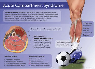
This educational medical illustration examines the anatomical and physiological basis of acute compartment syndrome within the lower leg. Through a detailed cross-sectional visualization of the calf compartments and supporting explanatory graphics, the poster demonstrates how elevated intracompartmental pressure impairs circulation, reduces tissue oxygenation, and can ultimately result in ischemic injury.
Created in 2025 using Procreate and Adobe Illustrator.
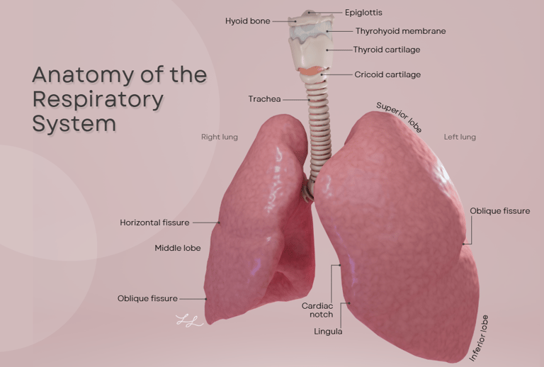
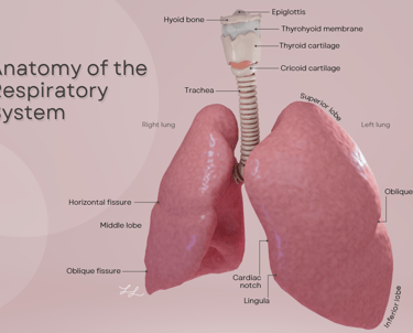
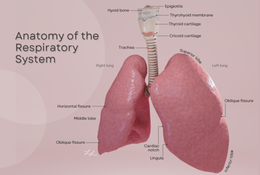
Anatomy of the Respiratory System
This educational medical illustration was developed to provide a clear anatomical overview of the upper and lower respiratory tract. The visualization highlights major respiratory structures, including the larynx, trachea, and lungs, while identifying key anatomical landmarks such as the pulmonary lobes, fissures, cardiac notch, and lingula.
Created in 2025 using Cinema4D, Redshift, and Adobe Illustrator
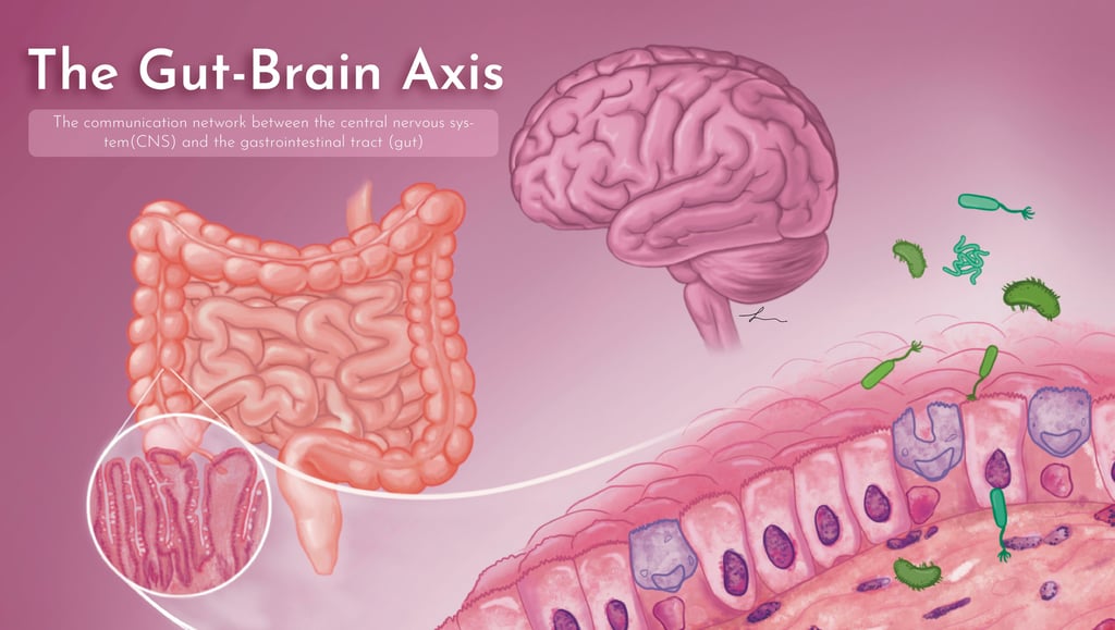
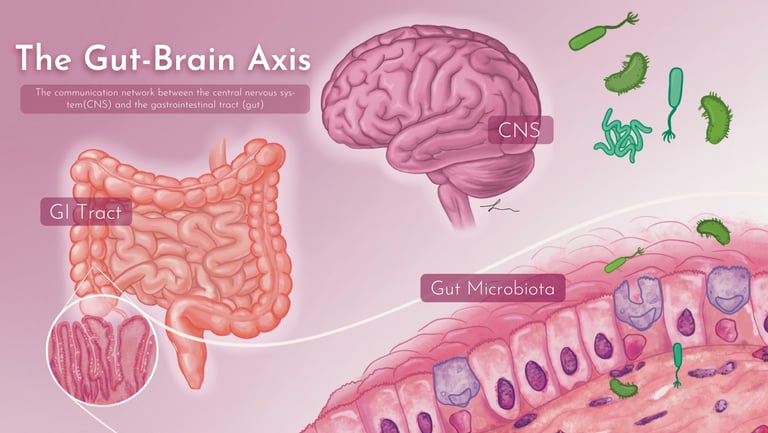
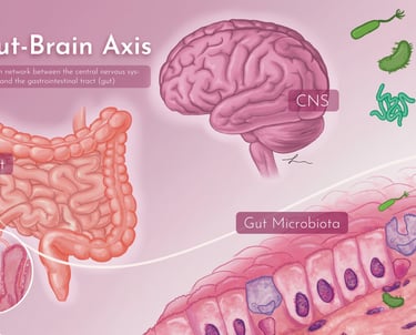
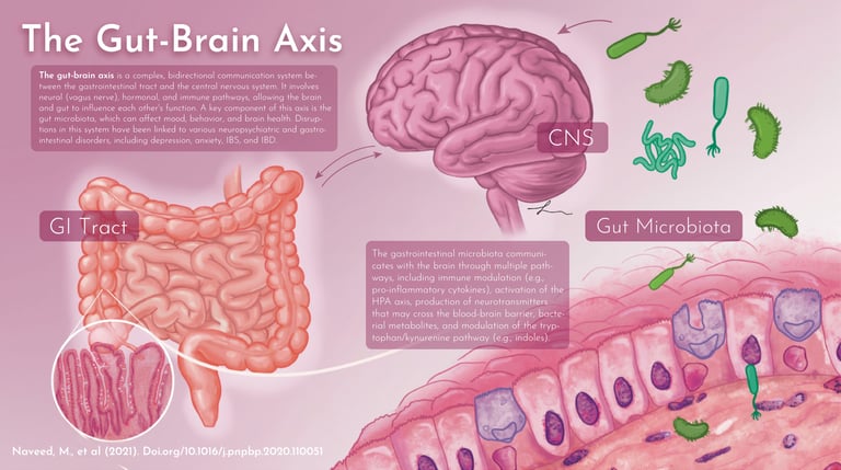
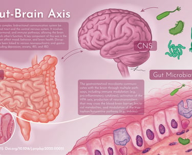
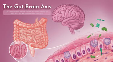
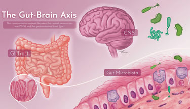
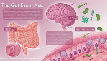
The Gut-Brain Axis: Biochemical and Neural Connection Network
Series of illustrations depicting the communication network between the CNS and GI system microbiota. Intended for Powerpoint presentation showing a title slide, then slides with more detailed descriptions and information.
Created in 2025 using Procreate and Adobe Illustrator.
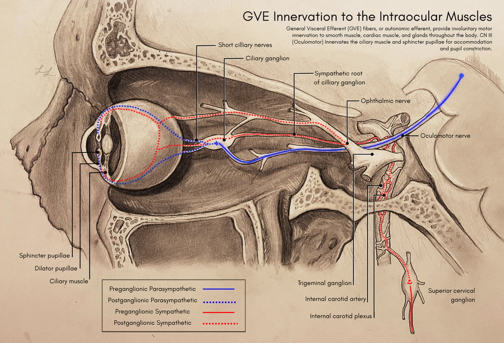
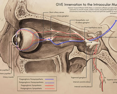
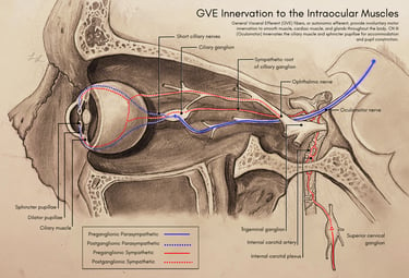
GVE Innervation Pathway to Intraocular Muscles
This piece depicts autonomic efferent innervation to smooth muscles of the eye, from preganglionic parasympathetic to postganglionic sympathetic. This piece was created using traditional drawing methods using graphite and paper, then brought into Adobe Illustrator for leader lines and labels.
Created in 2024 with traditional graphite and Adobe Illustrator
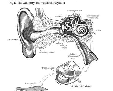
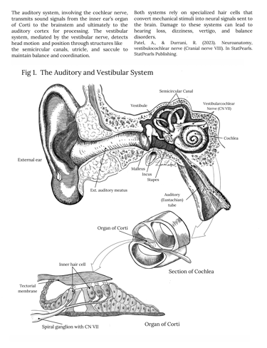
The Auditory and Vestibular System Textbook
This textbook-style medical illustration was developed to communicate the anatomy and function of the auditory and vestibular systems. The page combines a detailed overview of the external, middle, and inner ear with supplementary anatomical callouts of the cochlea and the Organ of Corti.
Designed to emulate professional anatomy textbook layouts, the project integrates scientific illustration, labeling hierarchy, and explanatory text to support student learning.
Created using Procreate and Adobe Illustrator.
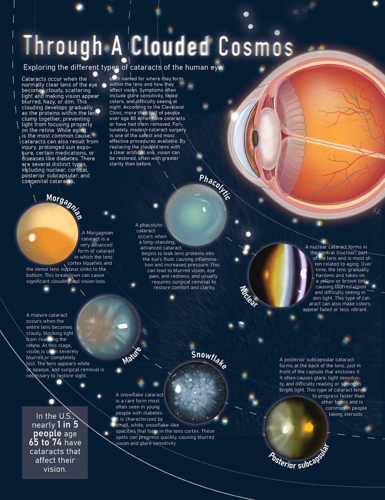
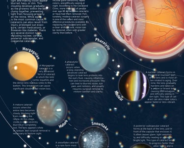
Through a Clouded Cosmos: Exploring Cataracts of the Human Eye
An educational ophthalmology infographic exploring the anatomy of the eye and the progression of multiple cataract subtypes. The piece guides viewers through several distinct cataract subtypes, including nuclear, posterior subcapsular, mature, Morganian, phacolytic, and snowflake cataracts.
Created using Procreate and Adobe Illustrator.
Through a Clouded Cosmos: Exploring Cataracts of the Human Eye

An educational ophthalmology infographic exploring the anatomy of the eye and the progression of multiple cataract subtypes. The piece guides viewers through several distinct cataract subtypes, including nuclear, posterior subcapsular, mature, Morganian, phacolytic, and snowflake cataracts.
Created using Procreate and Adobe Illustrator.
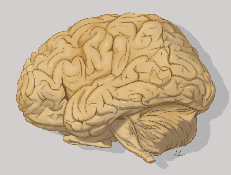
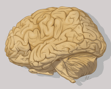
Brain Illustration
Detailed illustration of the human brain, highlighting sulci and gyri.
Created using Procreate
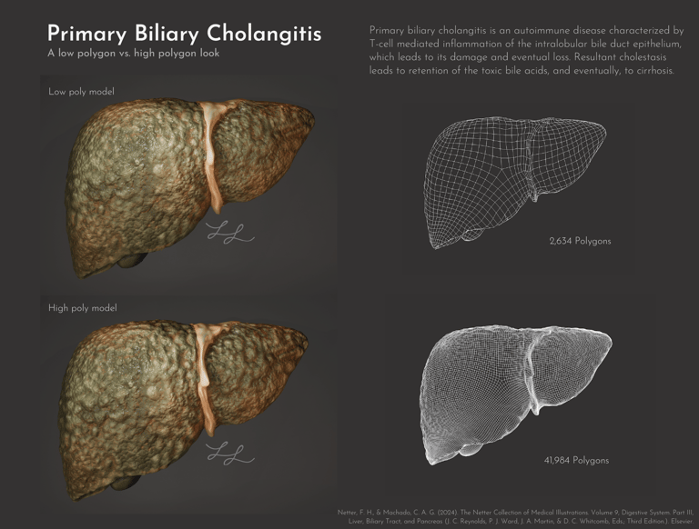
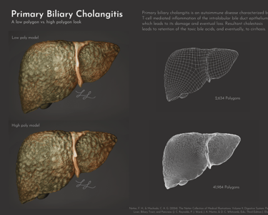
Primary Biliary Cholangitis: Pathological Liver Modeling and Topology Optimization
The project compares low-polygon and high-polygon modeling approaches, demonstrating the balance between detail and computational efficiency. Wireframe views are included to illustrate topology structure, polygon density, and optimization strategies used during model development. The final model was created to visualize the progressive destruction of intrahepatic bile ducts, resulting cholestasis, and eventual cirrhotic remodeling of the liver surface.
Created using Cinema4D, Redshift, and Adobe Illustrator.
Contact
Reach out for commissions or collaborations
contact
Copyright Lanford Biomedical © 2026. All rights reserved.