Medical Legal Visualization
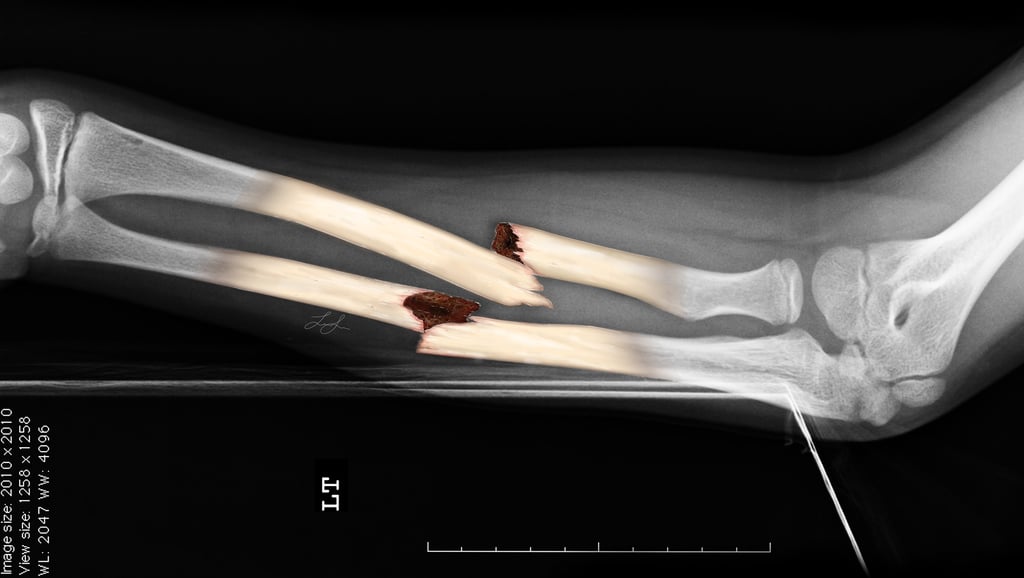
Medical-Legal Fracture Illustration: Displaced Radius and Ulna Fractures
Created from radiographic reference material, this illustration depicts displaced fractures of the forearm bones with emphasis on fracture morphology and alignment. The project required careful analysis of diagnostic imaging and the application of anatomical knowledge to reconstruct the injury in a visually accessible format.
Created in 2025 using Adobe Photoshop.
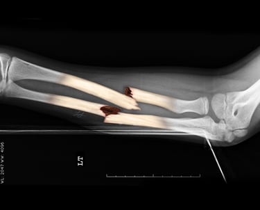
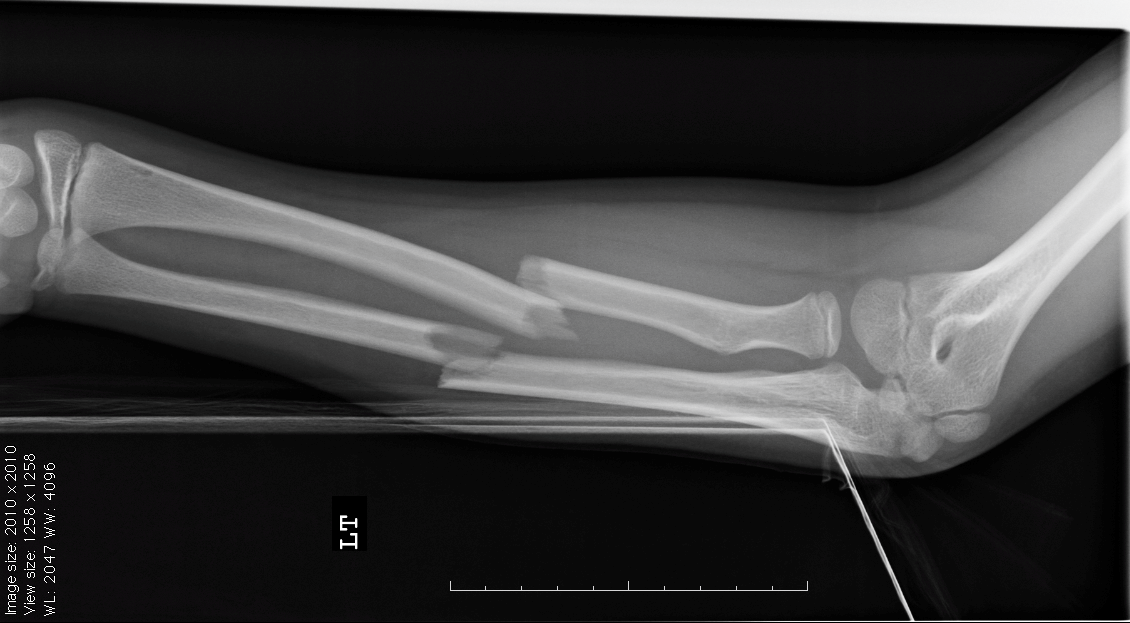
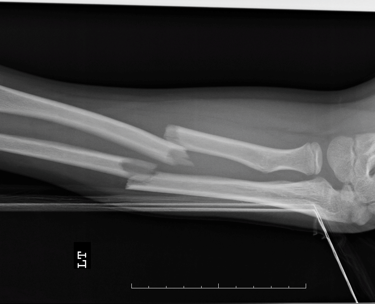
Original x-ray
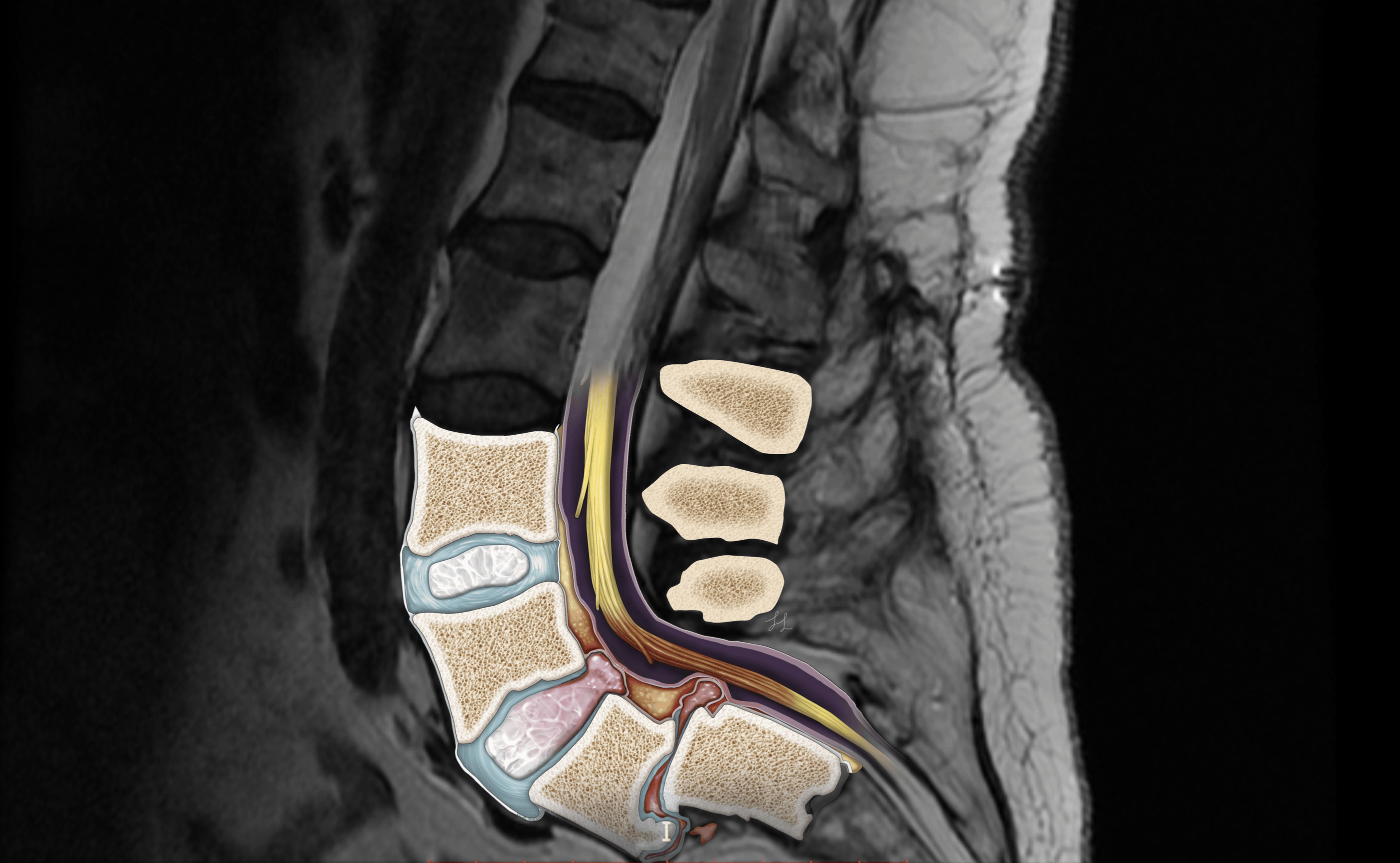
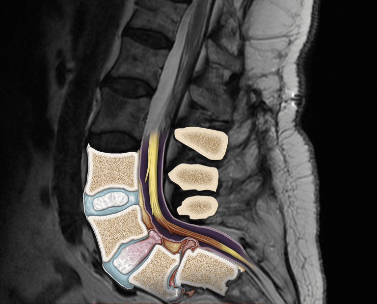
Lumbar Spine Injury Reconstruction from MRI Imaging
This illustration was developed from sagittal MRI reference images to visualize a traumatic lumbar spine injury and its effects on surrounding anatomical structures. The project required interpretation of radiographic findings and reconstruction of the affected vertebral bodies, intervertebral discs, spinal canal, and neural elements. By integrating diagnostic imaging with anatomical illustration, the final visualization clarifies the spatial relationships between the injury, spinal cord structures, and associated neural compression.
Created in 2025 using Adobe Photoshop.
Submammary Breast Augmentation: Surgical Procedure Illustration Series
This procedural illustration series visualizes the key stages of submammary breast augmentation, from preoperative marking and incision planning to implant placement and wound closure. The project was designed to communicate surgical workflow, anatomical relationships, and operative technique through a clear step-by-step visual narrative.
Created in 2025 using Procreate and Adobe InDesign.
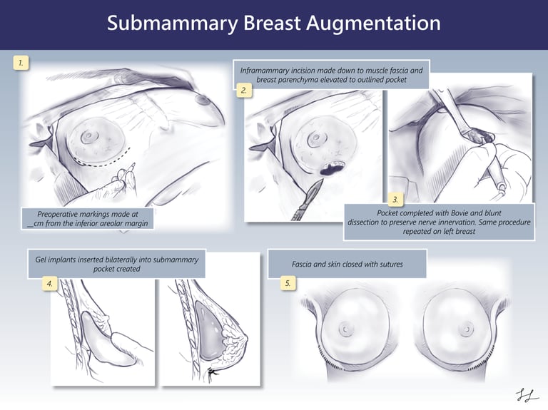
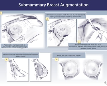
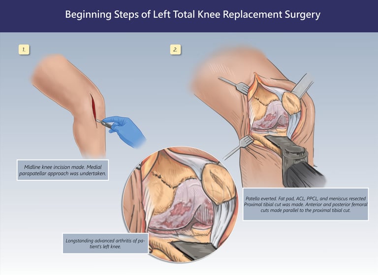
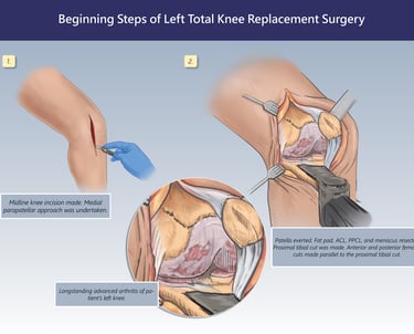
Exhibit 2: Intraoperative Vascular Injury and Repair
This exhibit focuses on a surgical complication that occurred during the arthroplasty procedure. Detailed illustrations demonstrate injury to the popliteal artery and vein, associated vascular damage, harvesting of the saphenous vein for grafting, and subsequent vascular reconstruction. The visualization was designed to clearly communicate the mechanism of injury and the corrective surgical intervention.
Created in 2025 using Procreate and Adobe InDesign
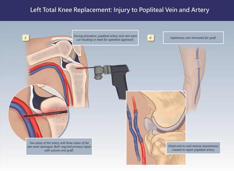
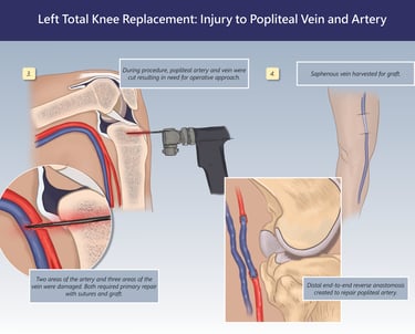
Exhibit 1: Initial Total Knee Replacement Procedure
This illustration depicts the opening stages of a left total knee arthroplasty, including the midline incision, medial parapatellar approach, joint exposure, and preparation of the femur and tibia for prosthetic implantation. Anatomical callouts highlight advanced degenerative joint disease and the surgical steps undertaken to restore joint function.
Created in 2025 using Procreate and Adobe InDesign
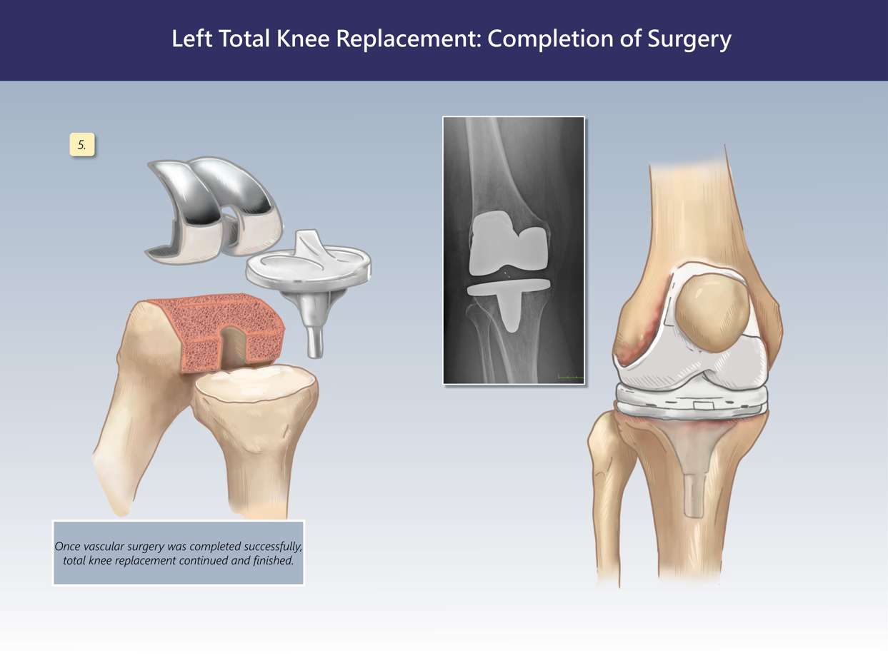
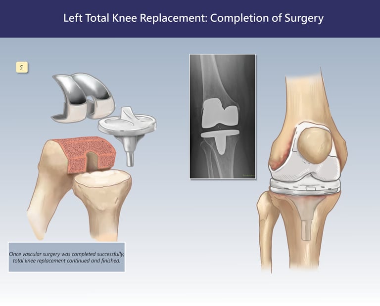
Exhibit 3: Completion of Total Knee Replacement
The final exhibit illustrates successful completion of the knee replacement following vascular repair. An exploded view of the prosthetic components, postoperative imaging, and final anatomical reconstruction demonstrate restoration of the joint and placement of the implant system.
Created in 2025 using Procreate and Adobe InDesign.