
Surgical Illustration
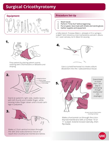
Steps to Perform Surgical Cricothyrotomy Infographic
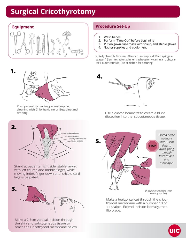
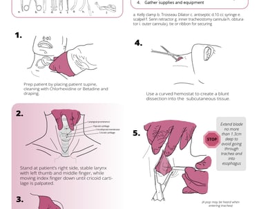
This is a two-page guide depicting the steps of how to perform a surgical cricothyrotomy procedure. This guide including the equipment needed, procedure set-up, and each step of performing the surgery.
Created in 2025 using Adobe Fresco and Adobe Illustrator.
Steps to Perform Surgical Cricothyrotomy Infographic
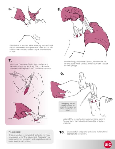
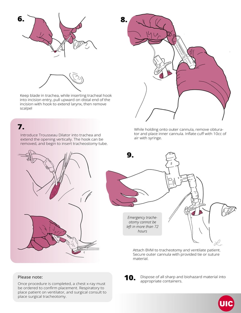
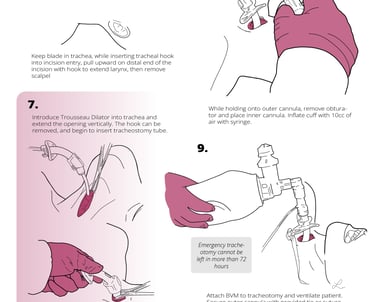
This is a two-page guide depicting the steps of how to perform a surgical cricothyrotomy procedure. This guide including the equipment needed, procedure set-up, and each step of performing the surgery.
Created in 2025 using Adobe Fresco and Adobe Illustrator.
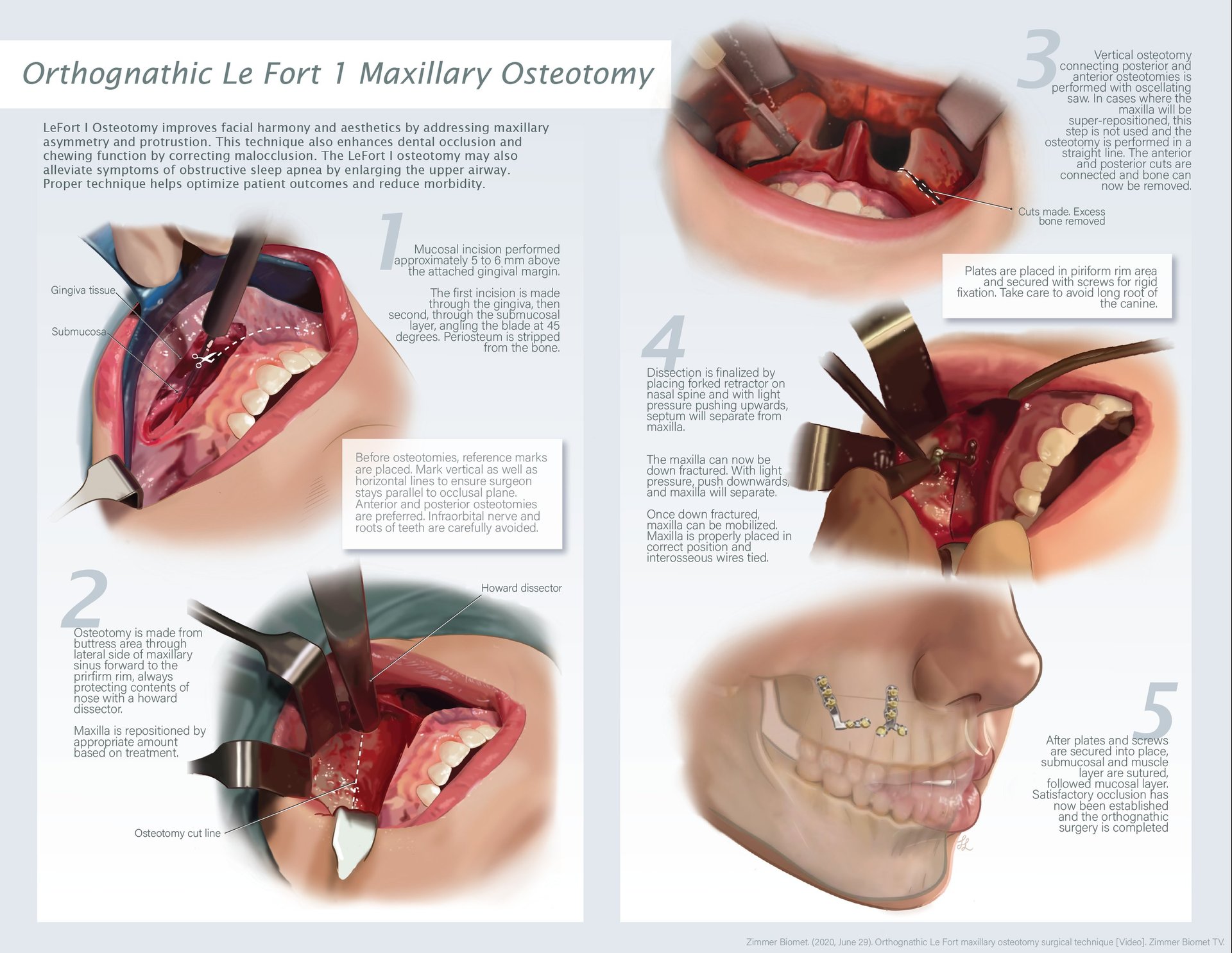
Orthognathic Le Fort 1 Maxillary Osteotomy: Steps for Procedure
This surgical illustration series depicts the key operative stages of a maxillary orthognathic Le Fort I osteotomy, a procedure commonly performed to correct dentofacial deformities and restore functional occlusion. Through five sequential illustrations, the spread visualizes critical surgical steps, including osteotomy placement, mobilization of the maxilla, repositioning of the skeletal segment, and stabilization of the corrected anatomy.
Designed for surgical education and clinical communication, the artwork combines anatomical accuracy with clear procedural storytelling to guide viewers through a complex craniofacial operation.
Created in 2026 using Procreate and Adobe Illustrator.
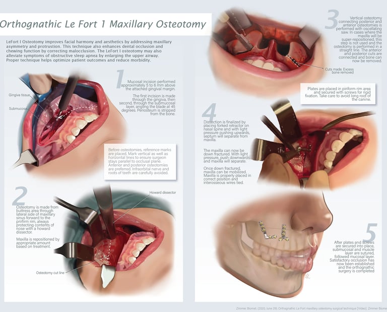
Orthognathic Le Fort 1 Maxillary Osteotomy: Steps for Procedure

This surgical illustration series depicts the key operative stages of a maxillary orthognathic Le Fort I osteotomy, a procedure commonly performed to correct dentofacial deformities and restore functional occlusion. Through five sequential illustrations, the spread visualizes critical surgical steps, including osteotomy placement, mobilization of the maxilla, repositioning of the skeletal segment, and stabilization of the corrected anatomy.
Designed for surgical education and clinical communication, the artwork combines anatomical accuracy with clear procedural storytelling to guide viewers through a complex craniofacial operation.
Created in 2026 using Procreate and Adobe Illustrator.
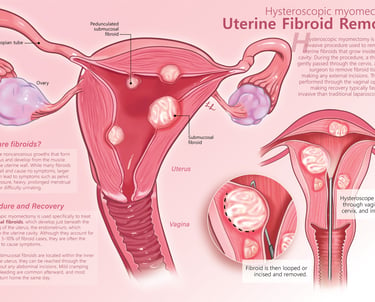
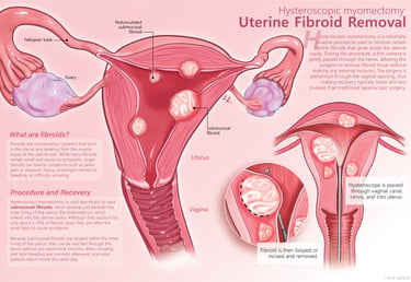
Uterine Fibroid Removal: Hysteroscopic Myomectomy
This educational surgical illustration combines anatomical and procedural visualization to explain the treatment of uterine fibroids through hysteroscopic myomectomy. The primary illustration presents a cross-sectional view of the uterus, emphasizing the relationship between fibroids and surrounding uterine anatomy. A secondary illustration demonstrates the minimally invasive surgical approach, showing the insertion of a hysteroscope and the resection of fibroid tissue.
Created in 2026 using Procreate and Adobe Illustrator.
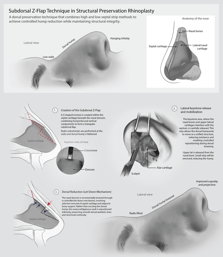
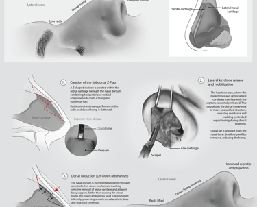
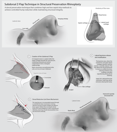
Structural Preservation Rhinoplasty: Subdorsal Z-Flap Technique
This educational surgical illustration was developed to explain a different preservation rhinoplasty technique through step by step illustrations. This piece focuses on clarity, anatomy, and education for surgical students/residents.
Created using Procreate and Adobe Illustrator.
Contact
Reach out for commissions or collaborations
contact
Copyright Lanford Biomedical © 2026. All rights reserved.